


Prelude
This May, the European Commission approved Amgen’s tarlatamab, a DLL3 × CD3 bispecific T-cell engager (TCE). This decision came after the DeLLphi-304 trial showed that tarlatamab extended median overall survival (OS) in second-line, extensive-stage small-cell lung cancer (SCLC) from 8.3 to 13.6 months. This result established tarlatamab as the first durable, second-line option in a tumor type that had resisted essentially every immunotherapy.
In the same month, Lilly reported Phase 3 data on retatrutide, a peptide drug that agonizes three surface receptors, GLP-1R, GIPR, and the glucagon receptor. It produced mean weight loss near 24% at one year, comfortably past tirzepatide and roughly twice what a pure GLP-1R agonist could deliver.
Finally, Merck and Kelun-Biotech reported that sacituzumab tirumotecan (sac-TMT), an antibody-drug conjugate (ADC) designed as a TROP2-directed antibody tethered to a topoisomerase 1 inhibitor, cut the risk of cancer progression or death by 65% versus pembrolizumab alone in first-line, PD-L1-positive advanced non-small-cell lung cancer (NSCLC) when added to pembrolizumab.
Despite engaging with different receptors and functioning through distinct mechanisms, these three compounds are deeply connected by the fact that they all leverage our rapidly growing understanding of cell surface dynamics.
Retatrutide activates three GPCRs that co-reside on pancreatic beta cells, integrating their signals into a single coherent effect. Tarlatamab binds DLL3, which is largely restricted to neuroendocrine tumor cells, forcing them into contact with T-cells by also engaging CD3 receptors. Sac-TMT leverages TROP2’s unique internalization route and a pH-sensitive bifunctional linker to deliver its cytotoxic payload to the lysosomes of tumor cells.
Over the last decade, our ability to interrogate cell surface dynamics has improved significantly as new measurement tools have emerged. The maturation of these experimental technologies alongside more advanced computational techniques will unlock a new wave of better-tolerated medicines with more precise and novel pharmacology.
The Cell Membrane and its Role in Drug Development
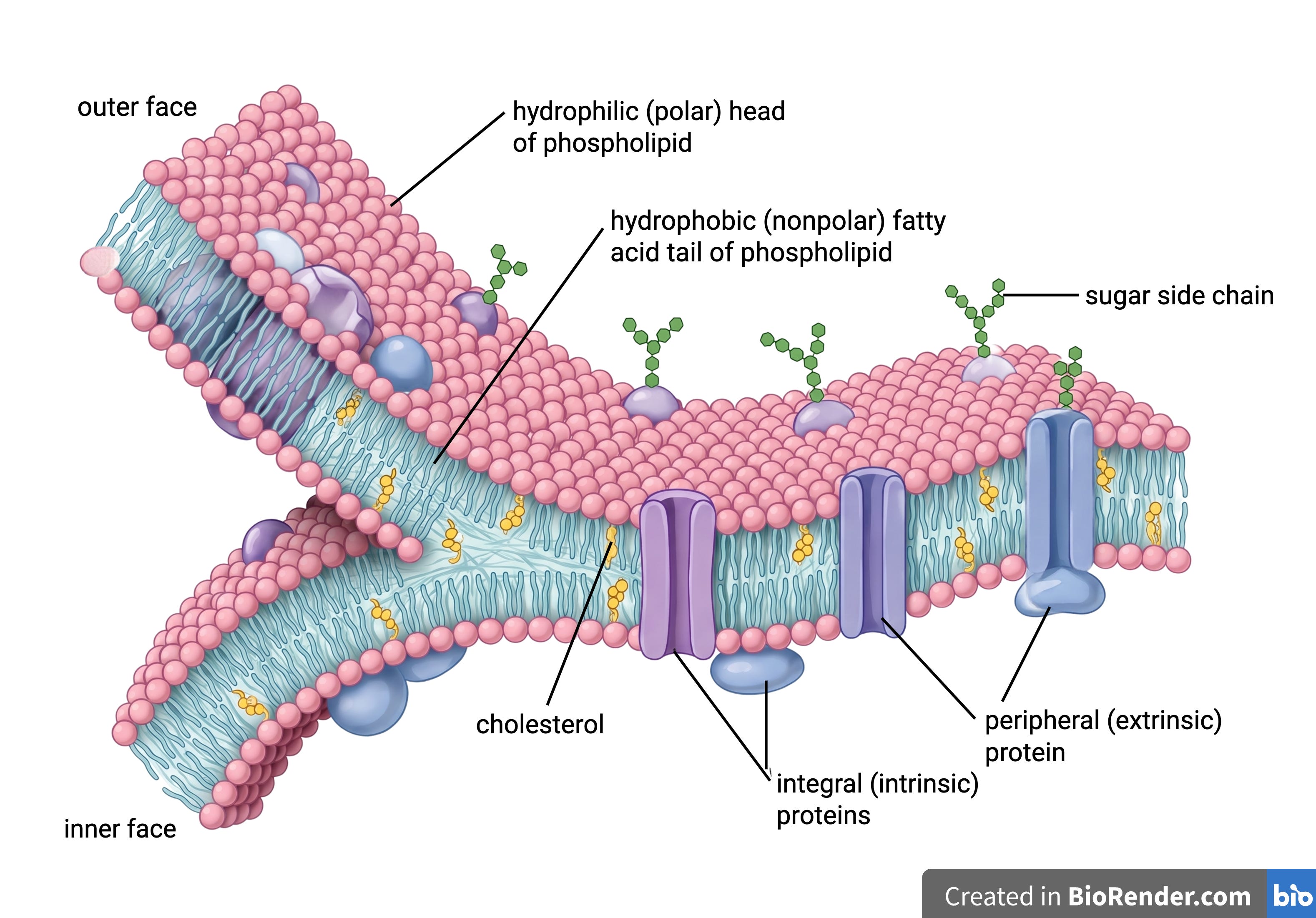
A cell’s surface (a plasma membrane composed of two layers of phospholipids) is a thin sheet about 5 nm thick on a cell 10–20 µm across, a roughly 1:3,000 ratio. The membrane is not bare: it is studded with proteins that serve as channels, pumps, receptors, and anchors that can move laterally through the lipid bilayer. A single cell carries on the order of 10⁶–10⁷ surface protein molecules drawn from 2,000–3,000 distinct species, packed tightly enough that neighbors sit ~6 nm apart. The two faces of the membrane differ in composition and are maintained as distinct compartments by dedicated enzymes, giving the surface a clear “inside” and “outside.”
The plasma membrane acts as the central hub coordinating cell-cell communication. Combined with its natural accessibility, the plasma membrane is a critical interface for drug development. Drugs can act on membrane proteins with exposed functional domains without crossing the bilayer, which blocks most large or polar drugs, like antibodies, from entering the cell. A receptor can also behave as a mechanical relay: binding on the outside changes conformation, clustering, or orientation of that receptor on the inside, triggering a biochemical cascade that elicits downstream intracellular signaling.
The membrane also offers routes inside via various pathways, including receptor-mediated endocytosis. A drug can bind a specific surface receptor and be carried into the cell as that receptor internalizes. Because this binding is selective, only cells displaying the target receptor take up the cargo, enabling precise, cell-type-specific delivery of otherwise impermeable payloads.
Arguably, no target has taught us more about the cell surface than HER2, at least not in such storied fashion. Its four-decade arc runs through all three of this essay’s questions: (1) what sits on the membrane and in what quantity, (2) what it sits beside, and (3) where it goes once a ligand binds it. The science has also advanced against an unusually dramatic backdrop, from an oncogene left in an abandoned freezer to patients who massed outside that company’s gates to demand access to a drug in early clinical trials.
Targeting the Surfaceome
None of the progress on HER2 would have happened if we were not able to resolve the cell surface with reasonable precision.
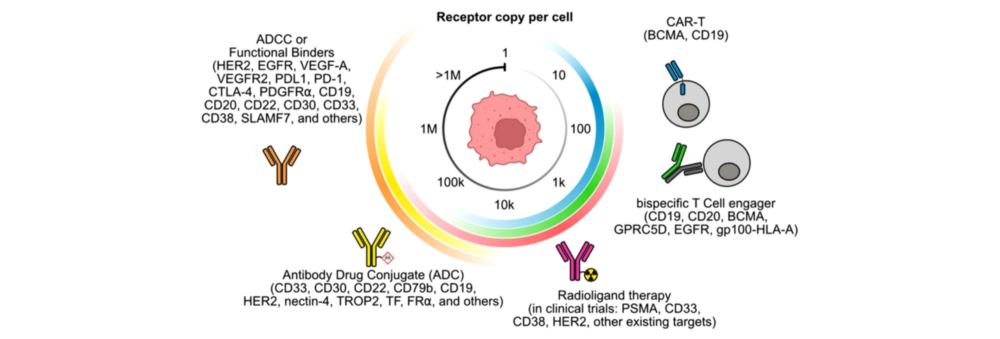
The surfaceome is the set of proteins exposed at the extracellular portion of the plasma membrane, plus the fraction of secreted and shed proteins that stay tightly cell-associated. There is no consensus on a definitive list of surface proteins and many proteins’ surface residency is context-dependent. Of the roughly 4,000-6,000 membrane and secreted proteins encoded in the genome, estimates show about half are expressed in any given cell. Moreover, the abundance of surface proteins spans across 5 orders of magnitude from over-expressed HER2 at 10⁶ copies per cell to an individual neoantigen on a cancer cell displayed at fewer than ten copies per cell.
Approved oncology drugs have targeted roughly 30. One reason for this small number is that cancer and healthy cells share many of the same surface proteins, making specificity difficult to achieve. Approved drugs therefore focus on targeting surface proteins that are more highly expressed in cancer cells as targeting non-specific proteins increases the risk of harming healthy cells. This difference in expression is the crux of therapeutic index.
In the nascent days of drug development, precisely identifying surface proteins was an insurmountable technical challenge. One promising solution was to leverage antibodies’ ability to specifically bind target proteins. By probing cell surfaces with known antibodies, scientists could deduce their surface-resident binding partners in a one-to-one manner.
Unfortunately, early production methods of antibodies used immunized animals so the antibody batches were polyclonal mixtures, heterogeneous serums consisting of thousands of different types of antibodies. Without knowing the identities of the probes, the cell surfaces could not be confidently mapped.
This ambiguity was resolved first in 1975, when César Milstein and Georges Köhler invented the hybridoma technique. Their breakthrough fused antibody-producing cells (B cells) taken from an immunized mouse with an immortal myeloma cell to create a cell line that secreted a single, monoclonal antibody, enabling researchers to raise antibodies against one cell surface protein at a time.
Armed with this tool to precisely isolate surface proteins, scientists turned their focus to understanding if certain proteins were differentially present on diseased cell populations, such as cancer cells. Such insights would potentially allow the development of precision therapy as opposed to a crude non-specific approach such as chemotherapy.
In the early 1980s, scientists at MIT pulled the neu oncogene out of chemically induced rat tumors. Neu encodes a large cell surface antigen structurally similar to the EGF receptor, a key protein involved in cell growth. A single valine to glutamate substitution in its transmembrane region made it oncogenic.
Later, at Genentech, Axel Ullrich’s group cloned the human counterpart, HER2. Dennis Slamon, one of Ullrich’s collaborators at UCLA, ran Ullrich’s probe against a San Antonio tissue bank. He found HER2 amplification, when cells express more of a protein than usual, in about 30% of 189 primary breast cancers, and that amplification independently predicted worse survival and earlier relapse.
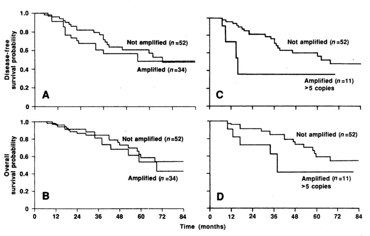
Since HER2 was substantially amplified on tumor cells and not normal cells, the target seemed to harbor exciting therapeutic potential. For strategic reasons, Genentech initially refused to fund the program because they had strong skepticism about the market and the efficacy of monoclonal antibodies for oncology. But an internal team kept it alive and produced muMAb 4D5, a mouse antibody that blocked the growth of cells overexpressing HER2.
The team then humanized it by grafting the six CDR loops of 4D5 onto a consensus human IgG1 framework. The final variant, humAb4D5-8, was a version with more than 250-fold greater affinity for HER2 in humans. This drove strong antibody-dependent cellular cytotoxicity, a phenomenon wherein target cells are marked for killing by the body’s immune cells.
That molecule became the storied trastuzumab. As it moved into trials, women with metastatic breast cancer who did not qualify still wanted it, but Genentech hesitated to release a scarce, experimental compound for compassionate use. Breast Cancer Action, founded by Elenore Pred, took up the fight. Marti Nelson, an advocate who was denied access to the drug, died in November 1994, galvanizing the movement further. On December 6 1994, patients and allies massed outside Genentech’s headquarters, demanding “access, not excuses.“ One protester asked, “How many women have to die on Genentech’s doorstep?“
The pressure worked. Genentech reversed course and invited advocates to help design the pivotal Phase 3 trials, the first partnership of its kind in the industry. It also opened a compassionate access lottery, which gave more than 300 women access to the drug.
The pivotal Phase III trial showed that adding trastuzumab to chemotherapy doubled response rates and improved OS in metastatic, HER2+ breast cancer. On September 25, 1998, the FDA approved Herceptin. The agency cleared it together with a companion diagnostic, Dako’s HercepTest, setting the pattern for every targeted therapy that followed.
Since the widespread adoption of trastuzumab, our ability to identify and quantify surface proteins has improved significantly. This is crucial, as surface residence is correlated with extracellular accessibility which is required by a standard antibody target.
Two complementary cell surface protein measurement families now dominate. The first and longest-established readout is liquid chromatography-tandem mass spectrometry (LC-MS/MS), an untargeted discovery method that analyzes dissociated, lysed cell mixtures.
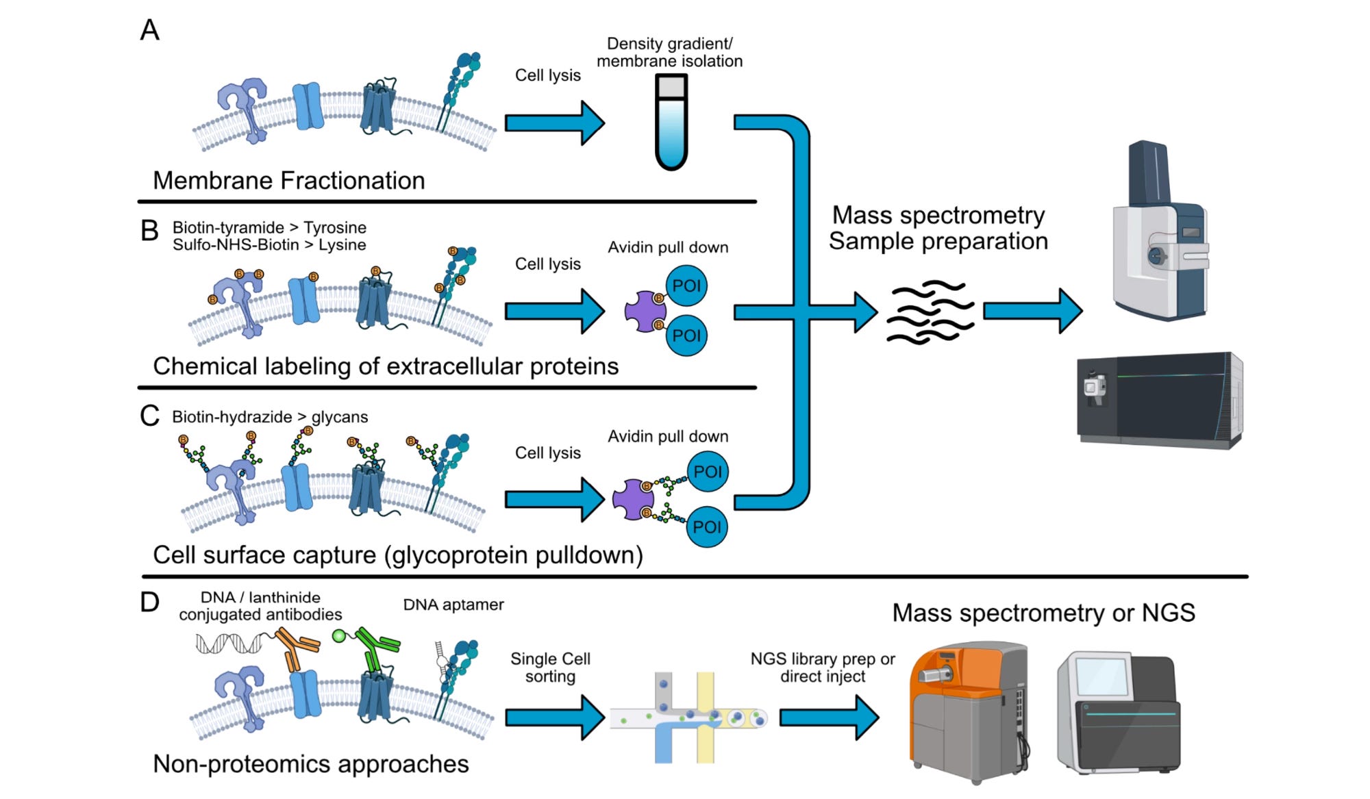
First, experimenters will run various sample preparation techniques, determining which proteins reach the instrument. Membrane proteins are roughly 100-fold less abundant than cell-internal proteins, so they are chronically undersampled in whole-cell lysate.
Cell-surface capture is one solution that works by tagging sugar molecules on the outsides of cells, enriching surface proteins at the same time. This method boosts surfaceome purity from roughly 10% in whole-cell lysate, to 40–60% with gentler membrane-tagging implementations, to about 95% with full glycocapture. Nevertheless, the stringent selection constrains output efficiency, meaning most MS runs may only recover about 200 total proteins per million input cells.
An acquisition scheme then chooses what proteins are detected by the MS. Data-dependent acquisition (DDA) selectively fragments precursors; data-independent acquisition (DIA) systematically scans fixed windows, and can now quantify 3,000–5,000 proteins per cell.
LC-MS/MS-based workflows have major limitations including low throughput at the single-cell level resolution. The low sensitivity of existing instruments means at most 1 out of 10,000 proteins is captured, or up to 5,300 protein molecules in a single cell. Moreover, deep label-free single-cell proteomics can only operate on the order of a few hundred cells per day. Comparatively, a routine scRNA-seq experiment profiles 10⁵ to 10⁶ cells.
Given the intrinsic sparsity of the protein capture process, higher throughput is necessary to increase the signal-to-noise ratio and provide a clearer view of surface protein distribution across cell types. The resulting measurement will improve confidence during target specificity assessment, modality selection, and on-target off-tissue toxicity effects prediction.
The second family is affinity-based profiling: a targeted approach that trades discovery for pre-validated binders against known epitopes. Two strong differentiators from this set of methods are its ability to target low-abundance proteins that may be missed by MS and to measure directly on intact tissues since this family typically uses fluorescent imaging probes that bind to barcoded antibodies.
The leading method is Bruker’s CosMx which collects multiomics data, pairing whole-transcriptome imaging with a 64-plex antibody protein panel on the same slide, at 0.0144 μm² per pixel. The breast cancer panel unsurprisingly contains HER2.
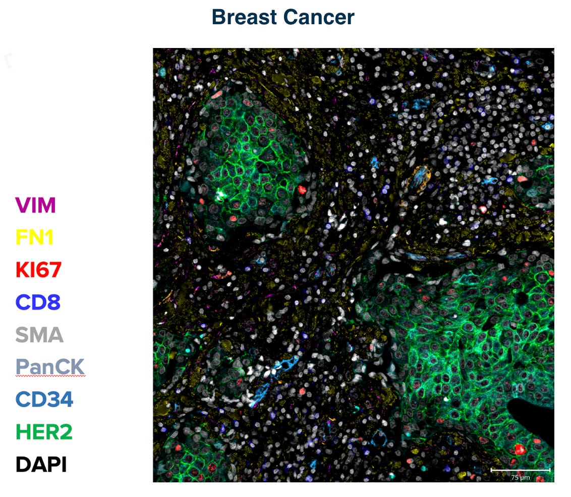
Subcellular resolution delivered through a commoditized, standardized workflow allows these tools to scale seamlessly, making them an attractive data-acquisition foundation for training tissue foundation models as demonstrated in a recent Noetik-Bruker collaboration.
The lack of a singular perfect experimental method combined with commercialized data acquisition workflow and emerging capability of computational inference to extract salient signal from high-dimensional data is a fertile ground for target discovery innovation. By curating a large collection of omics data and developing advanced machine learning algorithms, researchers can gain unique insights into cell-state-dependent surface protein expression. Consequently, researchers can prioritize a list of top-ranked combinations from a seemingly intractable number to develop surface engagers and depleters with improved precision and reduced side effects.
Cartography Biosciences is executing on this exact premise. The company has developed an annotated reference of millions of tumor and normal cells across tissue types to nominate novel target pairs. While its lead phase 1 asset, a LY6G6D × CD3 T-cell engager, was engineered to beat the failed linclatamig, Cartography’s target discovery engine has independently gained traction through a target-discovery collaboration with Pfizer, and a first target option exercised by Gilead in triple-negative breast cancer (TNBC). Epana Bio points the same logic at autoimmunity and builds precision autoimmune therapies aimed at the cellular origin of disease.
Unraveling the Proxiome
A single abundant and locally specific target is often insufficient for the development of a safe and efficacious drug. One more durable route to cell-type selectivity is achieved by pairing two or more individually non-selective antigens on the cell surface so that only cells displaying both are engaged. Think of this like an AND gate in computing. A result only should occur if conditions A and B are both met.
Selecting target pairs is a nontrivial task because the cell surface is crowded, with an average nearest-neighbor spacing of roughly 6 nm. The study of the spatial organization and interaction, the proxiome, examines the higher-order surface networks where interactions manifest through physical complex formation and functional coupling. The proxiome can be directly measured or inferred by layering on structural prediction and prior protein-protein interaction (PPI) knowledge.
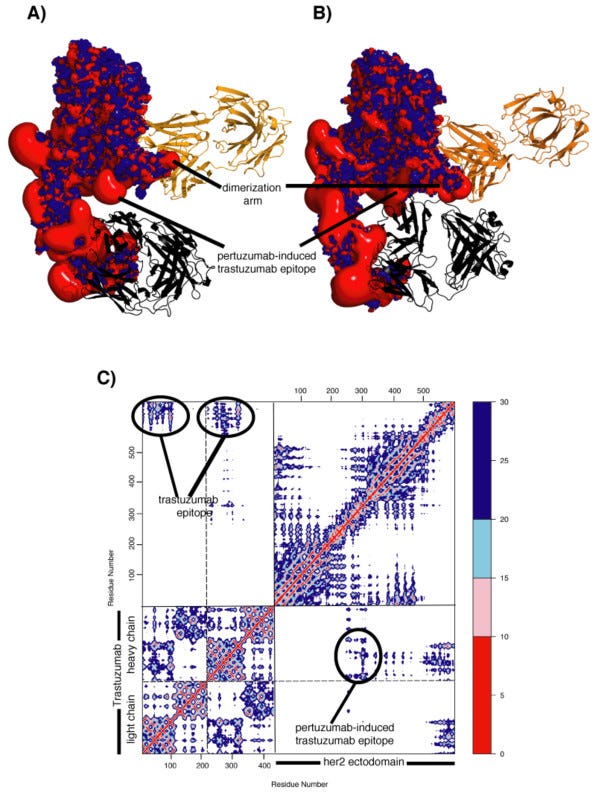
The classical gold standard approach for directly characterizing physical protein-protein interactions was targeted co-immunoprecipitation (Co-IP) which uses a specific antibody to capture a known “bait” protein and its interactors. The precipitate is then analyzed via Western blot, using an antibody against a suspected “prey” protein to confirm or deny the specific, hypothesized physical interaction between the targeted pair. In fact, this exact technique was applied to study HER2’s cell surface neighborhood, consequently leading to additional drug development.
The quintessential work was published in 1994 when scientists at Genentech demonstrated that HER2 pairs with HER3 to form the most potent growth signaling dimer in the family within a fibroblast-like cell line. With this novel insight, Genentech built pertuzumab, which binds the dimerization arm and blocks HER2 from partnering with HER3 and other HER-family receptors. Due to the complementary mechanism, trastuzumab and pertuzumab are often co-administered. In the landmark CLEOPATRA trial, adding the antibody combo to chemotherapy extended median OS to 56.5 months compared to 40.8 months with trastuzumab and chemotherapy alone.
While Co-IP can be very precise, the traditional workflow that depends on Western Blot limits the throughput to a single pair per experiment. Over the last decade, scientists have developed various labeling techniques to unveil the proxiome. These techniques utilize both MS-based (proximity labeling) and affinity-based (proximity ligation) workflows for more quantitative identification as we saw in the surfaceome case.
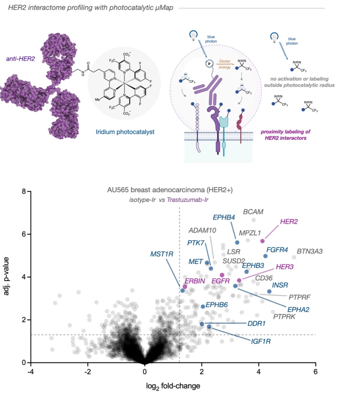
The state-of-the-art approach for proximity labeling, photocatalytic labeling, has become mainstream over the last 5 years. The mainstream leader in this family is μMap (micro-Map) which conjugates an iridium photocatalyst to a secondary antibody. Blue light drives Dexter energy transfer to convert diazirine into a carbene which has a short half-life of around 1 to 2 ns to enable a labeling radius of ~50 nm. A more recent approach, MultiMap, uses a single commercial dye, eosin Y, to activate four commonly used photoprobes to span multiple radii in one tunable workflow at a substantially lower cost than iridium.
μMap first mapped T-cell surface partners (CD45, CD29, CD47) and PD-L1 at the T-cell/target synapse. The same method was applied to the EGFR and c-MET interactomes. Last year, researchers profiled the HER2 interactome / proxiome across human breast cancer lines and identified galectin family proteins as uniquely enriched in trastuzumab-resistant models using μMap. Genetic and pharmacological galectin inhibition destabilized HER2 and other receptor tyrosine kinases, enhanced antibody-mediated receptor degradation, and restored trastuzumab sensitivity. They discovered hundreds of neighbors including HER3, providing a firmer mechanistic basis for the development of potential bispecific treatments around HER2.
In proximity ligation (PLA), two oligonucleotide-tagged antibodies are used to detect a protein pair within 50 nm. This approach trades the unbiased exhaustive characterization around 1 target for the preservation of spatial information and more targets of interest, potentially uncovering hundreds of unique PPIs at near single-cell resolution in one measurement. This integration helps distinguish interactions associated with specific functional cell states from proximity alone and, when coupled with perturbation, disentangle their downstream mechanisms.
The leading academic work is Sprox-seq, which can profile 528 pairwise surface PPIs in addition to mRNAs. The commoditization of a related approach is spearheaded by Moleculent whose offering features a smaller panel.
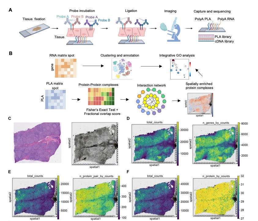
Recent technology development on labelling chemistry and probe design made the technology more suitable for obtaining insights from human tissues. μMap-FFPE and Sprox-seq can both run on formalin-fixed, paraffin-embedded (FFPE) patient blocks which make up more than 90 percent of the archived tissue samples available for biomedical research in the United States. Nevertheless, the translational difficulty is exacerbated by our limited ability to extract salient signals from highly heterogeneous samples.
Disease-state surface biology shifts with inflammation, hypoxia, prior therapy, stromal remodeling, and tumor evolution, so a target pair that looks selective on a tissue microarray can fail in minimal residual disease or in post-treatment relapse. How we can establish a causal link between a disease-associated surface PPI and disease phenotypes in a precise manner remains a huge bottleneck.
Proximity labeling’s strength lies in its ability to exhaustively search the neighborhood around a well-validated target. InduPro published their informatics workflow leveraging μMap as a target identification engine via spatial protein proximity. Using graph-based machine learning approaches on their proximity and expression data primarily derived from healthy and cancerous cell lines, they identified CDCP-1 to be an EGFR protein interactor.
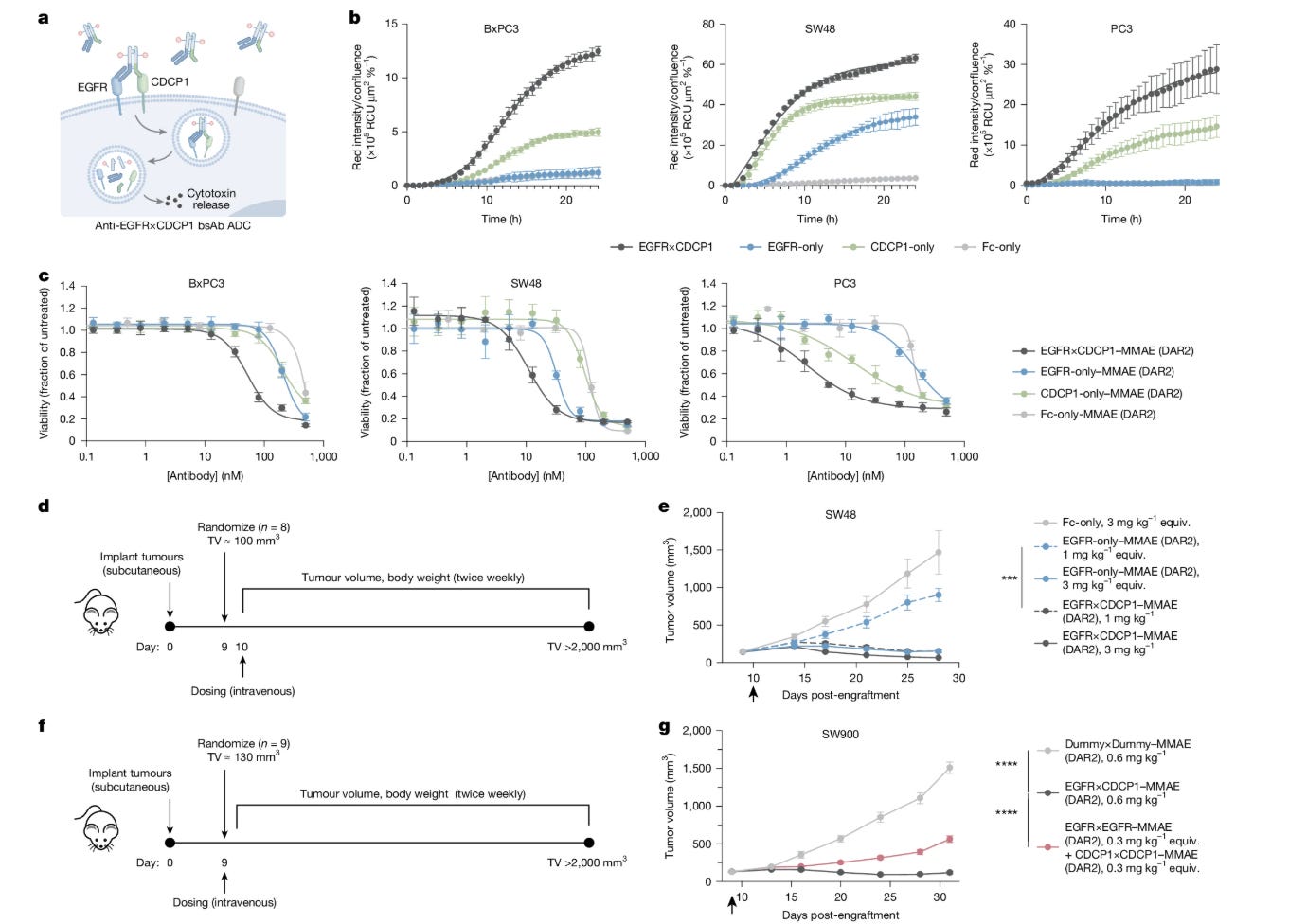
The company then screened more than 250 bispecific formats and ranked them on internalization and cytotoxicity, arriving at an EGFR domain III arm paired with a membrane-distal CDCP1 arm for their bispecific ADC program.
The rationale for an EGFR×CDCP1 bispecific ADC predates modern proximity mapping. More than a decade ago, researchers showed that CDCP1 forms ternary complexes with Src and EGFR via Co-IP, promoting oncogenic signaling. EGF activation also stabilizes CDCP1 and promotes its recycling to the cell surface, establishing a trafficking connection relevant to ADC delivery. Nevertheless, these insights are arguably far from mainstream. InduPro’s work increased the confidence of this local association across more in vitro and in vivo models, in addition to human tissues. InduPro further established the key mechanistic insights via isogenic CDCP1 knockouts.
InduPro’s results also suggested EGFR-HER3 to be strongly associated on the cell surface, providing some mechanistic insights on Izalontamab Brengitecan, the first approved bispecific ADC targeting EGFR-HER3 following June approval in China. The InduPro target discovery engine further supplied the immunology leg through a Sanofi collaboration on a bispecific PD-1 agonist for autoimmune disease and a multi-target oncology partnership with Lilly worth up to roughly $950M in milestones.
Drug development can leverage both sets of tools due to their complementarity. For example, PLA is more suited for discovering novel biology and for patient stratification, while proximity labeling is a natural fit for guiding multi-specific biologics design. These observations can be used to identify missing interactions in established models and to guide reconstruction of better in vivo and in vitro models, enabling the assessment of mechanism through genetic perturbation.
Elucidating surface residence and neighborhood compositions with mechanistic insight provides the baseline for feasible targets. But the interaction mode between a molecule and the surface protein dictates the possible mechanism of action and the therapeutic ceiling.
For instance, CD3, the T-cell target of conventional TCEs, illustrates how sophisticated molecular engineering can establish a therapeutic class. The T-cell receptor–CD3 complex can internalize upon engagement. Most modern TCEs therefore incorporate lower-affinity CD3-binding arms, which can reduce internalization, allowing TCEs to remain at the cell surface and bring T cells and tumor cells together for efficient killing. By contrast, therapies designed to deliver a payload inside the cell such as ADCs and siRNA conjugates require uptake and routing to the right intracellular compartment. Understanding these routes requires separate measurements.
Traversing the Deliverome
The deliverome focuses on the study of the lifecycle of internalization, from routing through a trafficking pathway to releasing into the right compartments of the right cell in the right tissue.
The science of receptor-mediated endocytosis matured in the 1990s. Alexander Sorkin studied how ligand binding drives EGFR internalization and subsequent degradation. By combining EGFR’s extracellular domain with HER2’s intracellular domain, his team showed that the resulting chimeric receptor internalized less efficiently than EGFR, suggesting that HER2’s intracellular machinery was less suited to rapid uptake.
But uptake is only part of the story. Tracking a bound ligand can be misleading: how quickly it dissociates determines how faithfully it follows the receptor, rather than taking a separate intracellular route. Later imaging of HER2-bound trastuzumab revealed another important distinction: the antibody entered endosomes, compartments that sort incoming cargo, but was efficiently recycled to the surface rather than sent to lysosomes, the acidic, enzyme-filled compartments that break cargo down. HER2 was therefore not simply resistant to internalization; efficient recycling was responsible for limited intracellular accumulation.
Despite the internalization-resistant nature, HER2 sits at high density on tumor cells, so even modest internalization can carry enough payload inside for cell killing. With trastuzumab’s clinical and commercial success, Genentech viewed HER2 as an attractive delivery target, paving the way for the development of Genentech’s first ADC, trastuzumab emtansine, sold as Kadcyla and approved by the FDA in February 2013. Conjugated via a noncleavable linker, the payload releases only after the cell fully breaks the antibody down in the lysosome, with limited cleavage in the bloodstream, where free payloads would contribute to chemotherapy-like toxicity. In the EMILIA trial, it lifted median OS from 25.1 to 30.9 months against lapatinib plus capecitabine.
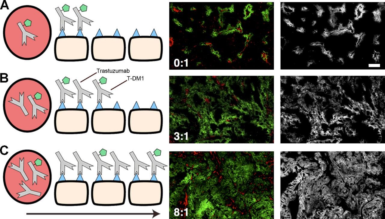
Unfortunately, Kadcyla’s strong dependence on HER2 expression left out the majority of patients. This accessibility issue is further compounded by the tumor’s poor drainage which restrains antibodies’ movement through dense stroma largely by diffusion. The fraction that arrives is not spread evenly as in vitro studies showed that Kadcyla dosed at clinically relevant levels occupies a perivascular rim and leaves antigen-positive cells deeper in the tissue untouched, with penetration recovered only by co-administering unconjugated trastuzumab to compete for the binding sites nearest the vessel.
Daiichi Sankyo’s trastuzumab deruxtecan, sold as Enhertu, went further by pairing trastuzumab with a cleavable linker which enables extracellular cleavage in the tumor microenvironment. This engineered bystander effect let Enhertu reach tumors that Kadcyla could not. The FDA first cleared it in December 2019 on DESTINY-Breast01, in later-line HER2-positive disease.
The more consequential moment came in August 2022, when DESTINY-Breast04 nearly doubled progression-free survival in HER2-low breast cancer. That approval roughly tripled the number of patients who could benefit from targeting HER2, and the label extended again to HER2-ultralow in January 2025.
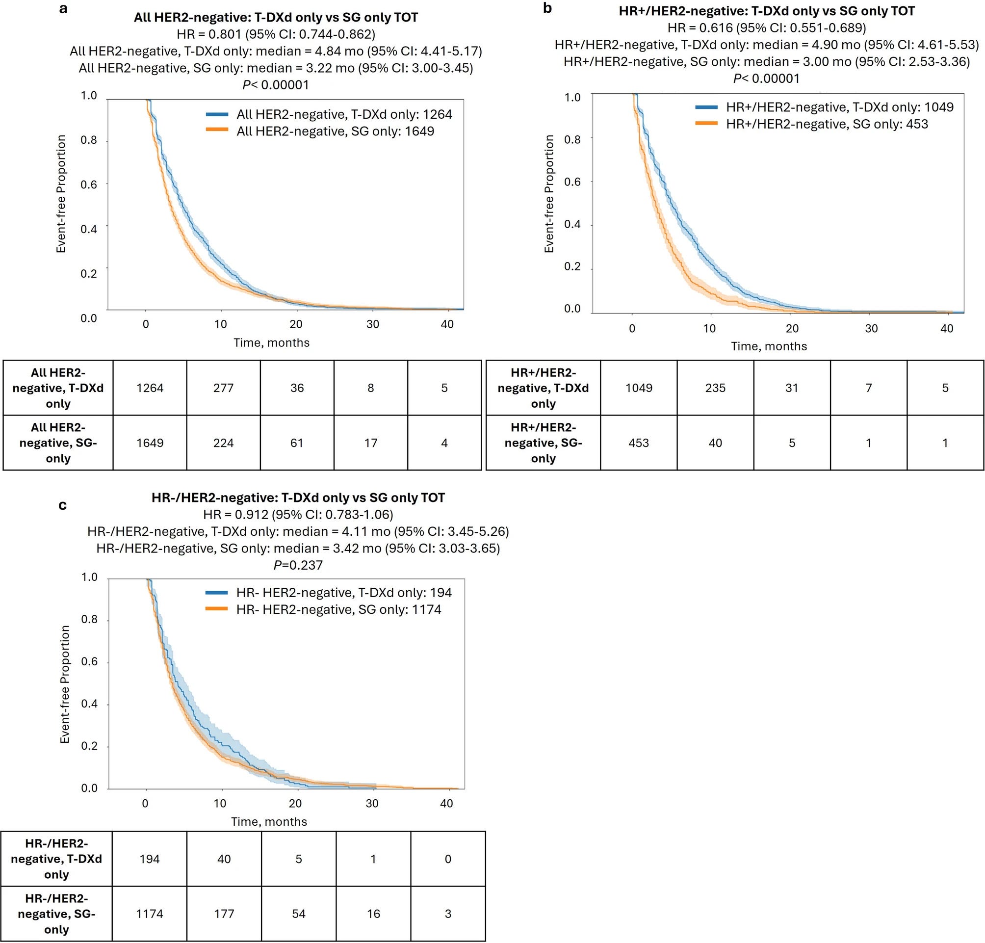
Unfortunately, the cleavable linker of Enhertu is a double-edged sword. Its chemical predisposition to cleavage leads to more respiratory adverse effects including interstitial lung disease (ILD) as HER2 is also lowly expressed in healthy lung cells. Recent progress has centered around the Trop-2 target with the approval of Trodelvy and Sac-TMT which has a cleaner safety profile for lung-related side effects but higher risk for neutropenia. Notably, neutropenia is easier to manage than ILD for clinicians.
But the prospect on efficacy is currently unclear; a meta-analysis comparing Enhertu and Trodelvy within the HER2-negative population showed that the HER2-targeting drug has a significantly prolonged time on treatment outside of TNBC tumors. Partial trial results from Sac-TMT on HR+/HER2- and TNBC are out; head-to-head statistical analysis in the near future will provide more clarity for patients and clinicians.
Despite their recent successes and momentum, ADCs still have a long way to go especially with respect to potent delivery. Review estimates place less than 1% of an administered ADC at the tumor site, and under 0.01% of the injected dose in each gram of tumor. That number does not get much prettier for other modalities either. For siRNA, endosomal escape into the cytosol occurs at roughly 1–2% efficiency. Delivering a larger fraction of each dose to the desired cells would let developers lower doses and reduce the systemic exposure that drives off-target toxicity.
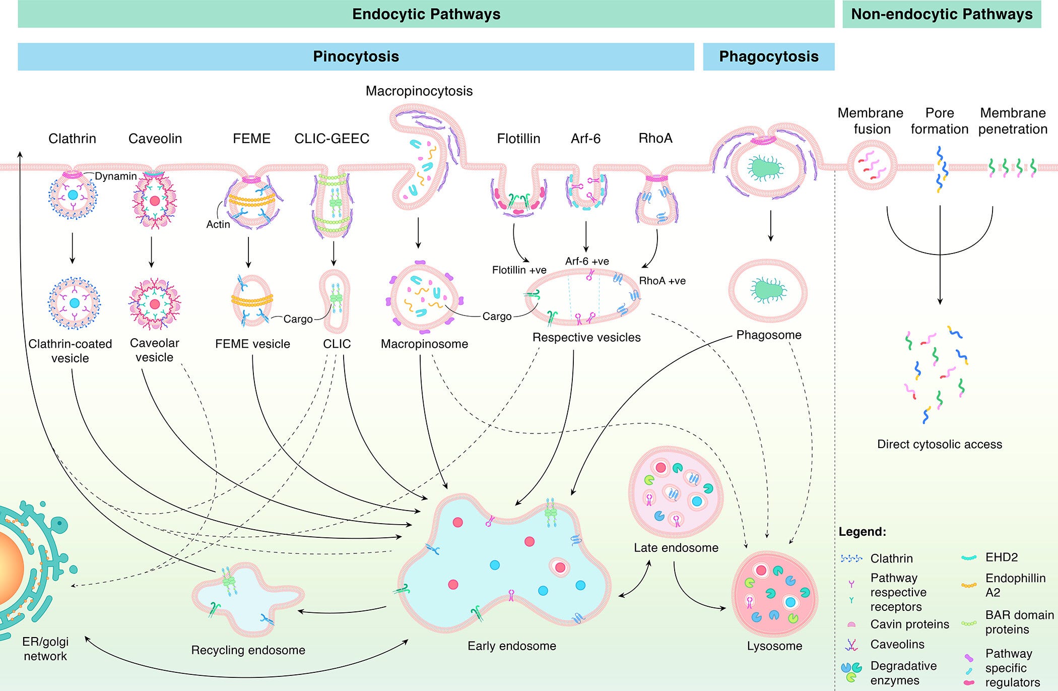
As our ability to design molecule candidates continues to improve, higher throughput approaches for identifying candidates that have good biodistribution and internalization mechanisms will be crucial for rational drug discovery. For ADCs, matrix-assisted laser absorption ionization imaging has been used to map the distribution of released payload, supported by MS for quantification at the tissue level.
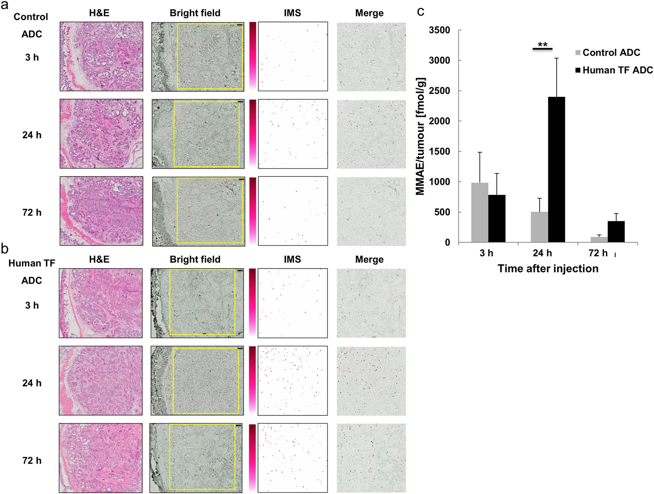
Dissecting the fate of a payload at a sub-cellular resolution requires different approaches. In a reductionist model, receptor-mediated endocytosis carries a drug into an endosome after binding. What must happen next depends on the payload: macromolecules such as siRNA generally need to escape into the cytosol before lysosomal degradation, whereas many ADCs are intentionally routed to lysosomes, where the harsh conditions help release the small-molecule payload that can then diffuse or be transported across the lysosomal membrane into the cytosol.
A standard commercialized approach tracks uptake into the endocytic compartment. It uses pH-activated dyes which stay dark outside the cell or inside the cytosol and switch on once the receptor carries them into an acidified vesicle, turning internalization into a readout on a high-throughput well plate. The dye is typically conjugated at a lysine or cysteine residue, restricting the method to protein-based therapeutics. Since the environment becomes progressively more acidic from endosome to lysosome, various dyes have been designed to light up at different stages.
Adding a cathepsin-activated probe in a second channel separates early internalization from lysosomal degradation. For conjugates whose linkers are cleaved by cathepsins, the probe is correlated with payload release. For siRNA, quantifying cytosolic delivery directly is more tractable as live-cell imaging can resolve single endosomal escape events at single-cell resolution.
To further resolve distribution at a compartment-specific level, nanoscale secondary ion mass spectrometry (NanoSIMS) is used. This technology detects isotopic labels directly. Oligonucleotides can incorporate brominated nucleotides during synthesis, while antibodies can be labeled by growing producer cells in heavy nitrogen. Each candidate requires custom production, which makes this the highest-resolution view of intracellular distribution available, but at the lowest-throughput.
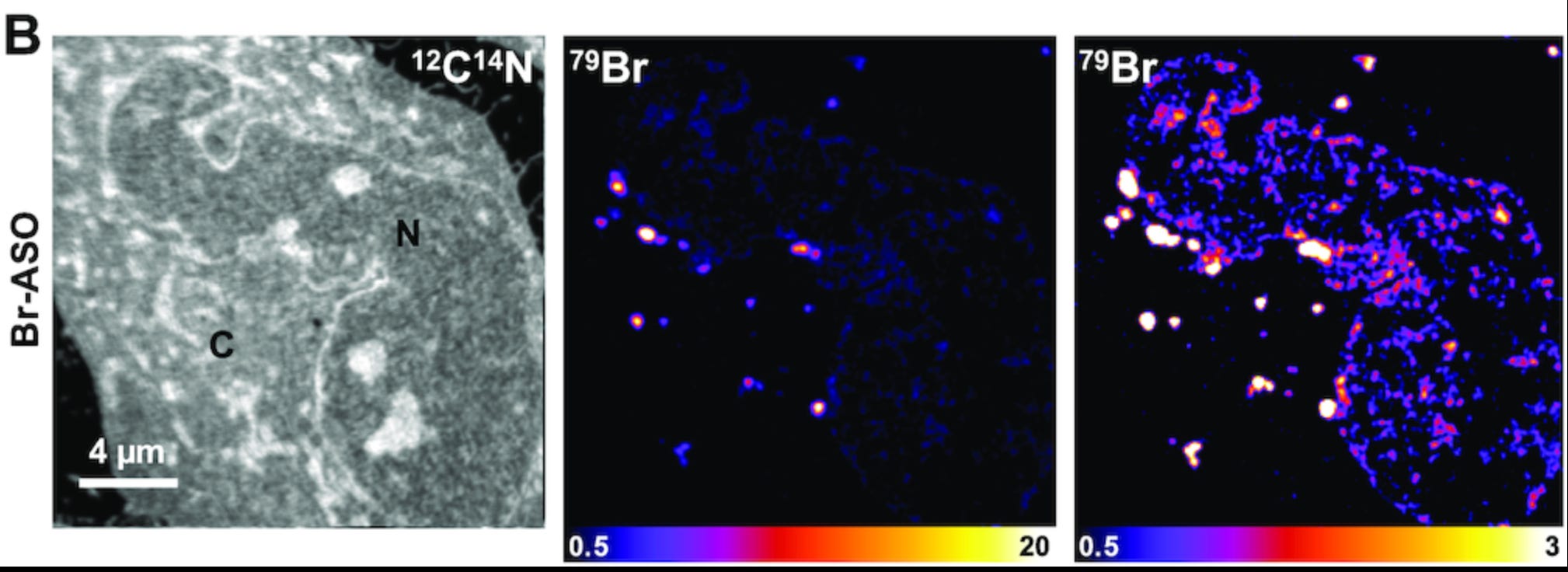
These innovations in measuring biodistribution at varying resolution can help drug developers to assess location-specific release with more certainty.
Recall an earlier point on the difficulty of circumventing the dense tumor stroma; an alternative strategy is to approach the stroma as a depot rather than a barrier. This can be achieved by directing a non-internalizing ADC at a structural scaffold (like collagen IV) or a matrix-anchored matricellular protein (like tenascin-C); promising activity has been shown in preclinical models.
One front runner in industrializing ADC is Ridge Bio. Its ProTrigger linkers are ML-designed, protease-cleavable prodrug systems intended to activate in the tumor microenvironment or stroma, with the stated aim of unlocking extracellular and poorly internalizing targets for conjugates while reducing off-tumor toxicity.
On the siRNA side, Nosis Bio is focused on unlocking extra-hepatic delivery via machine learning-enabled protein design. Its Connexa platform combines a proprietary atlas of cell-specific internalizing receptors with AI-powered molecular design and high-throughput in vivo characterization to develop receptor-targeted delivery vehicles.
Innovations from companies will primarily accumulate as proprietary knowledge. Therefore, systematic and centralized open-source experimentation holds outsized value for drug discovery. The Deliverome Project, an early-stage FRO, is positioning to perform those foundational measurements by standardizing datasets for internalization, escape, and tissue uptake, potentially compressing early-stage discovery by roughly a year per program.
Human translation of measurements remains a huge bottleneck. Absolute internalization kinetics differ significantly between in vitro and in vivo across both ADCs and siRNA conjugates. Nevertheless, the rank order tends to persist reasonably well. The ADC literature reaches a similar conclusion: across 19 ADCs, in vitro and in vivo tumor-static concentrations correlate at a Spearman ρ of 0.82, with in vivo values roughly 27-fold higher in absolute terms. The magnitudes are wrong but the rank order persists, making a screen useful in a relative sense.
Unfortunately, this reassurance has boundaries. The evidence sits mostly in vitro to xenograft rather than mouse to human, and it does not generalize across modalities as demonstrated in a head-to-head comparison of hundreds of nanoparticles for which in vitro and in vivo delivery poorly correlated.
The Convergence of Technologies Can Accelerate the Development of Safe and Efficacious Medicines
The greater opportunity lies in the convergence of all three sub-domains: by interlinking measurement technologies to decipher a cell’s surface and where a bound molecule travels, we have a systematic basis for translating surface biology insights into medicines with wider therapeutic windows and novel mechanisms of action.
The near-term trend will likely center on sophisticated molecular engineering of bispecifics around a validated target to include a less validated but promising target. InduPro’s EGFRxCDCP1 is a great example illustrating this approach with EGFR commanding 5 FDA-approved antibody inhibitors and CDCP1-directed ADCs (e.g., HY0001) still only in early trials.
The more ambitious long-term opportunity is to adopt a similar framework for discovering novel pharmacology that leverages surface interactions to reshape associated cellular circuits.
One such exciting area is the modulation of innate immune cells in immune-mediated diseases. Currently, all approved immune cell engager therapies for cancer are exclusively T-cell engagers which can induce adverse cytokine release syndrome which is an intrinsic risk due to CD3 engagement. Despite tumor-associated myeloid cells being the most abundant immune population in many solid tumors, accounting for up to half of the tumor mass, classic innate checkpoint inhibitors targeting the CD47/SIRPα axis have largely failed in the clinic.
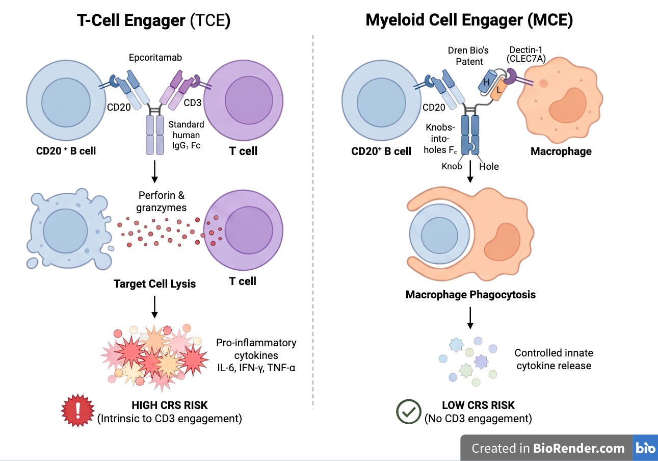
Myeloid engagers are changing the paradigm. Dren Bio and LTZ Therapeutics both build molecules that recruit myeloid cells through a phagocytic receptor and kill cancer cells via antibody-dependent cellular phagocytosis and tumor microenvironment reprogramming. Dren’s Dectin-1 x CD20-directed myeloid engager DR-0201 (acquired by Sanofi) is in trials in B-cell lymphoma and in a basket of autoimmune rheumatic diseases, and DR-0202 is in a phase 1 trial for a basket of solid tumor indications. LTZ Therapeutics is also innovating in this area. Their phase 1 lead asset, LTZ-301, is selectively depleting pathogenic CD79b-positive B cells via an Fc-gamma receptor-independent mechanism with pre-clinical programs focusing on solid tumors.
Even though the role of Dectin-1 in antifungal defense and tumorigenesis was explored in academic settings, it was under-studied relative to its adaptive immune cell counterparts. The improvement in measurement technologies will unveil more targets like Dectin-1 and greatly accelerate target validation efforts with more direct human evidence that can guide design of appropriate pre-clinical models.
Beyond recruiting immune cells, better maps of the cell surface could reveal new ways to reshape disease niches and direct immunity toward previously overlooked disease targets.
Tertiary lymphoid structures (TLSs) offer one application. Their stromal organization and lymphotoxin-dependent development suggest testing a locally anchored organizer-receptor agonist to strengthen antitumor immunity, or a localized maintenance blocker to suppress a pathogenic autoimmune niche.
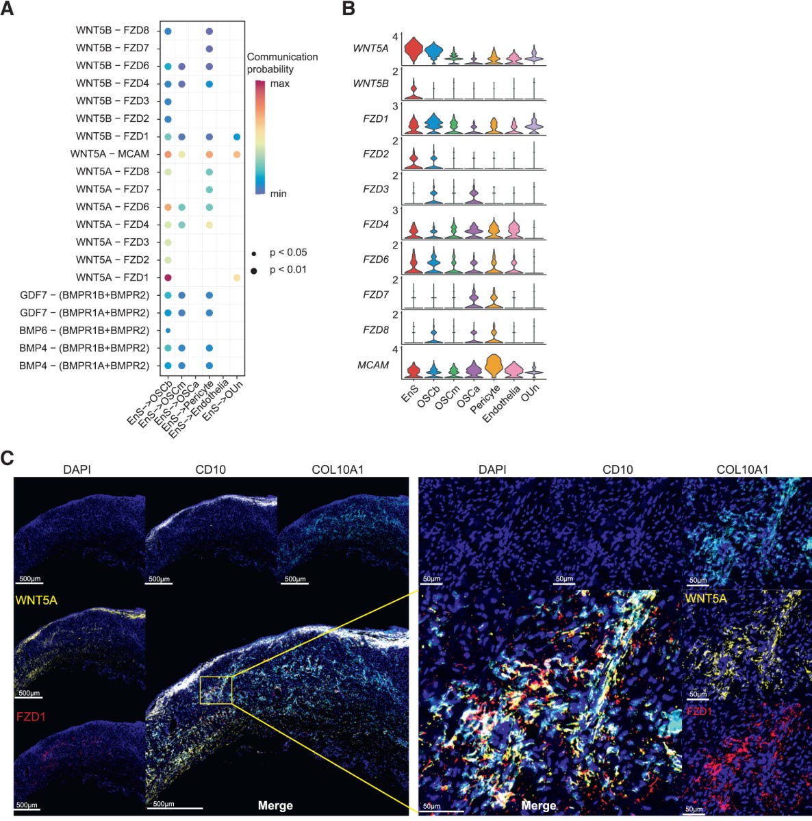
Endometriosis is another debilitating underserved disease. Early works are looking into the niche interactions that sustain lesion growth; by identifying pathogenic surface combinations that spare normal cyclical endometrium, potential therapeutics can selectively remove a validated pathological population.
Another area where this convergence could shine is neoantigen-targeting cancer vaccines, an asset class that has finally gained deserved hype again after pessimism that lasted over a decade. Merck and Moderna recently reported that intismeran autogene with pembrolizumab met recurrence-free and distant-metastasis-free survival endpoints, the first positive Phase 3 result for its class.
For an mRNA cancer vaccine to be efficacious, the mRNA must enter antigen-presenting cells and reach the cytosol, where it is translated into antigen proteins. These cells break the proteins into peptides and display them on their surface using human leukocyte antigen (HLA) molecules which help activate associated T cells and drive their expansion. The resulting cancer-targeting CD8 T cells must then reach and enter the tumor. Crucially, cancer cells must also process their own antigen proteins and display matching peptides on HLA class I molecules. Otherwise, the primed T cells cannot recognize and kill them.
For intismeran autogene, tumor and matched-normal DNA sequencing, tumor RNA sequencing, and HLA typing from every patient are used to guide the mRNA design which encodes up to 34 neoantigens, a limit set by delivery and manufacturing concerns.
When I worked at Moderna, I learned the difficulty of selecting the right targets as only 0.5% of non-silent mutations are presented as peptide-MHC complexes. Moreover, an individual neoantigen species may be displayed at fewer than ten copies per cell, against a total surface peptide-MHC load on the order of 10⁵ complexes.
Precisely measuring peptides on the surface of tumor cells may therefore greatly improve vaccine efficacy. Recent efforts measure these peptides directly using mass spectrometry. An mRNA vaccine designed this way achieved strong responses in glioblastoma bone marrow-derived macrophage co-cultures.
As such measurement technology and the ability to assess mRNA endosomal escape continue to improve, discovering the holy grail, an off-the-shelf neoantigen cancer vaccine, may be closer to reality than we think.
Conclusion
HER2 took four decades to teach us important lessons. Abundance and relative specificity gave trastuzumab its window, a neighbor gave pertuzumab its mechanism, and where the molecule went after binding gave Kadcyla and Enhertu theirs. These novel treatments collectively turned HER2 over-expression from a poor prognosis to a sign of hope for a longer life for patients.
What has changed since the early 2000s is that all three of surfaceome, proxiome, and deliverome now have commoditized measurement technologies. And these technologies will greatly facilitate the rational design of medicines with reduced side effects, improved efficacy, and exploit novel pharmacology to benefit underserved patients within the next decade.
Postlude
For clarity and focus, we omitted many interesting topics, including glycosylation and the extracellular matrix in basic science, as well as genetic medicine, in vivo CAR-T, and other therapeutic modalities. We also did not discuss delivery-vehicle engineering including antibodies, peptides, and nanoparticles which is a complementary key component in designing next-generation therapeutics.
Disclosure
Dimension has invested in Epana Bio and Ridge Bio.
Acknowledgment
I sincerely appreciate the expertise and feedback from
Becca Carlson (co-CEO, Deliverome Project)
Huili Wang (Postdoc, UChicago)
Feng Yi (CSO, KeLun)
Kang Yong Loh (Postdoc, Arc institute)
Joey Federico (PhD Candidate at PME, UChicago)
Haotian Du (Protein Designer)
Jeff Blethen (CEO, Stealth Biotech)
Yuchen Liu (PhD Candidate in Public Health, Harvard)
Junjie Xia (Computational Biologist, Broad Institute)